Home
/ Pelvic Anatomy Ligaments, Schematic Illustrations Of The Pelvic Ligaments Muscles And Nerve Download Scientific Diagram, It is close to the major vasculature of the pelvis, including external iliac vein.
Pelvic Anatomy Ligaments, Schematic Illustrations Of The Pelvic Ligaments Muscles And Nerve Download Scientific Diagram, It is close to the major vasculature of the pelvis, including external iliac vein.
Pelvic Anatomy Ligaments, Schematic Illustrations Of The Pelvic Ligaments Muscles And Nerve Download Scientific Diagram, It is close to the major vasculature of the pelvis, including external iliac vein.. Broad ligament the broad ligament supports the uterus, fallopian tubes, and ovaries. The pelvis's frame is made up of the bones of the pelvis, which connect the axial skeleton to the femurs, and therefore acts in weight bearing of the upper body. The ligaments of the pelvis, are amongst the strongest in the human body. Female pelvis ppt by mayil rasamani), which are reflections of the broad ligament attaching the ovaries to the lateral pelvis. Other ligaments attached to bony pelvis include the sacrococcygeal ligaments, pubic symphysis ligaments, and endopelvic fascia ligament.
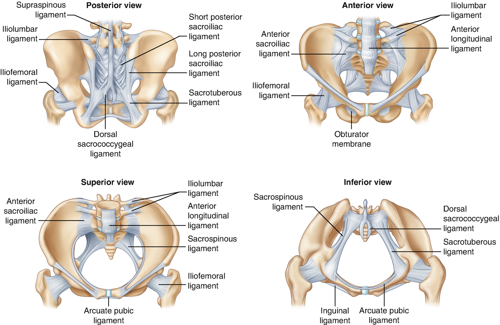
This is the final level of the pelvis that baby descends and rotates through during birth. • with intraperitoneal fluid collections, ligaments will appear moderately thin and hyperechoic. The pelvis's frame is made up of the bones of the pelvis, which connect the axial skeleton to the femurs, and therefore acts in weight bearing of the upper body. The vertebropelvic ligaments include the iliolumbar, sacrotuberous and sacrospinous ligaments. We hypothesized differences in the strength of various pelvic ligaments and therefore, aimed to evaluate and compare their biomechanical properties.
1 from The vertebropelvic ligaments include the iliolumbar, sacrotuberous and sacrospinous ligaments. These are accessory ligaments to the sacroiliac joints of the pelvis and are important in maintaining its stability. It is close to the major vasculature of the pelvis, including external iliac vein. Cookies allow us to analyze and store information such as the characteristics of your device as well as certain personal data (e.g., ip addresses, navigation, usage or geolocation data, unique identifiers). The floor of the pelvis is made up of the muscles of the pelvis, which support its. The ligaments of the pelvis, are amongst the strongest in the human body. citation needed this facilitates reconstruction of the floor of the inguinal canal. The inlet to the pelvic canal is at the level of the sacral promontory and superior aspect of the pubic bones.
We hypothesized differences in the strength of various pelvic ligaments and therefore, aimed to evaluate and compare their biomechanical properties.
The inlet to the pelvic canal is at the level of the sacral promontory and superior aspect of the pubic bones.; The pelvis is a boney structure at the base of the lumbar spine. The outlet is formed by the pubic arch, ischial spines, sacrotuberous ligaments, and the coccyx. Ligaments connect one bone to another and provide important stability. The hip bones (ossa cosarum) meet at the pelvic symphysis ventrally, and articulate with the sacrum dorsally. The 3 groups of ligaments are: We hypothesized differences in the strength of various pelvic ligaments and therefore, aimed to evaluate and compare their biomechanical properties. The outlet is formed by the pubic arch, ischial spines, sacrotuberous ligaments, and the coccyx. Like those osseous structures, the ligaments. This is part of the forced closure method that the pelvis adopts in order to keep itself secure. The pelvis consists of two innominate bones and the sacrum to which coccyx is attached. Those that connect the ilium to the sacrum; The named ligaments of the pelvis mostly arise from the sacrum and attach to varying segments of the pelvic bone.
Inherent stability of the pelvis is provided by ligaments. The ilium, ischium and the pubic bone. The pelvic girdle, also known as the hip bone, is composed of three fused bones: The ligaments that connect the sacrum to the coccyx. Pelvic ligaments • not routinely visualized by ultrasound.
1 from The inlet to the pelvic canal is at the level of the sacral promontory and superior aspect of the pubic bones.; Female pelvis ppt by mayil rasamani), which are reflections of the broad ligament attaching the ovaries to the lateral pelvis. It is close to the major vasculature of the pelvis, including external iliac vein. This is the final level of the pelvis that baby descends and rotates through during birth. The pelvis itself is a paired composite structure made up by three bones (ilium, ischium and pubis) that articulates with the sacral part of the axial spine. The broad ligament is a flat sheet of peritoneum, associated with the uterus, fallopian tubes and ovaries. Below the pelvic brim), posterior. The bottom level of the pelvis is the pelvic outlet;
This post discusses the anatomy of the pelvic ligaments, their structure, attachments, and how they mature through the decades of a person's life.
The pelvis consists of two innominate bones and the sacrum to which coccyx is attached. This post discusses the anatomy of the pelvic ligaments, their structure, attachments, and how they mature through the decades of a person's life. The 3 groups of ligaments are: Bones and ligaments of the female pelvis. The outlet is formed by the pubic arch, ischial spines, sacrotuberous ligaments, and the coccyx. The pelvis's frame is made up of the bones of the pelvis, which connect the axial skeleton to the femurs, and therefore acts in weight bearing of the upper body. This will be explored further on. The ligaments that connect the sacrum to the coccyx. The outlet is formed by the pubic arch, ischial spines, sacrotuberous ligaments, and the coccyx. The major osseous structures of the pelvis are wrapped in a complex fascial structure. Lets get deeper into the musculoskeletal anatomy of the hip and look at the bones and bony bits of the pelvis, and the ligaments that attach here and hold it. It is close to the major vasculature of the pelvis, including external iliac vein. The pelvis is the lower portion of the trunk, located between the abdomen and the lower limbs.
The pelvis is a boney structure at the base of the lumbar spine. Pelvic ligaments • not routinely visualized by ultrasound. The ligaments that connect the sacrum to the coccyx. Additional ligaments may be found in the female pelvis. The pectineal ligament is usually around 6 cm long in adults.
Anatomy Of The Female Pelvis Springerlink from media.springernature.com Inherent stability of the pelvis is provided by ligaments. The ligaments that connect the sacrum to the coccyx. Like those osseous structures, the ligaments. Imaios and selected third parties, use cookies or similar technologies, in particular for audience measurement. It extends to both sides of the pelvic wall. The pelvis's frame is made up of the bones of the pelvis, which connect the axial skeleton to the femurs, and therefore acts in weight bearing of the upper body. The 3 groups of ligaments are: It is close to the major vasculature of the pelvis, including external iliac vein.
The outlet is formed by the pubic arch, ischial spines, sacrotuberous ligaments, and the coccyx.
The pelvic outlet has two main ligaments that help support the ability of the sacrum to move out of the way to make more room forward/backward in the outlet. It is usually divided into two separate anatomic regions: The broad ligament is a flat sheet of peritoneum, associated with the uterus, fallopian tubes and ovaries. The ilium, ischium and the pubic bone. Ligaments connect one bone to another and provide important stability. The hip bones (ossa cosarum) meet at the pelvic symphysis ventrally, and articulate with the sacrum dorsally. The bony pelvis is held together with the support of the 3 vertebropelvic ligaments. These are accessory ligaments to the sacroiliac joints of the pelvis and are important in maintaining its stability. The 3 groups of ligaments are: The ligaments of the pelvis, are amongst the strongest in the human body. Cookies allow us to analyze and store information such as the characteristics of your device as well as certain personal data (e.g., ip addresses, navigation, usage or geolocation data, unique identifiers). It extends to both sides of the pelvic wall. The major osseous structures of the pelvis are wrapped in a complex fascial structure.